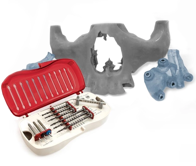
Atrophic maxilla is a condition characterized by a loss of bone volume in the upper jaw. It poses significant challenges in maxillofacial surgery and can affect facial aesthetics, overall oral function, and even psychological well-being.
However, today, advancements in dental surgical techniques provide innovative solutions to this significant dental condition. Notable procedures like the Zygomatic and Pterygoid implants, which are placed in zygoma (cheekbone) bones, or pterygoid plates (part of the sphenoid bone at the back of the upper jaw), are used in such conditions.
In this blog, we’ll explore how these bones play crucial roles in addressing atrophic maxillas, their anatomical importance, surgical implications, and therapeutic benefits.
Atrophic Maxilla-Definition and Understanding
The maxilla is the bone that makes up the upper jaw, cheeks, and lower parts of the eye sockets. Patients may present either after long periods of maxillary denture wear or following loss of the dentition related to severe periodontal issues. Severe atrophy of the maxilla hampers implant placement to allow restoration of the arch.
Atrophic maxilla arises from factors like tooth loss, periodontal disease, trauma, or congenital defects. It leads to the reabsorption of the alveolar ridge and subsequent bone loss. This condition makes traditional dental implant placement difficult, necessitating alternative methods to restore function and aesthetics.
How Zygoma bone acts as a structural pillar for implants
The zygoma, or cheekbone, is a key anatomical feature of the face. The zygoma is a very dense bone that provides excellent support for dental implants which can even replace a complete arch at once.
Its strong structure and strategic location make it ideal for supporting dental implants in severe maxillary atrophy cases. Introduced by Branemark in the 1990s, Zygoma implants are anchored directly into the zygomatic bone, detouring around the deficient maxillary ridge.
The Techniques Involved in Zygoma Implant and Considerations
Placing zygoma implants requires careful planning and surgical skills. Using Cone-beam computed tomography (CBCT) imaging for guided implant planning helps in preoperative assessment and planning. This technology precisely determines the zygoma’s anatomy and bone density. The surgical approach typically involves accessing the lateral area, avoiding vital structures like the infra-orbital nerve and maxillary sinus.
Therapeutic Advantages of Zygoma Implants
Zygoma implants offer a range of benefits in atrophic maxilla cases.
No Bone Grafting: They eliminate the need for extensive bone augmentation procedures, reducing surgical complexity and treatment time.
Stable and Strong: Anchored in dense zygomatic bone, these implants provide exceptional stability and support.
Immediate loading: This procedure allows immediate loading in some cases due to its exceptional density and strength. This speeds up functional restoration and increases patient satisfaction.

How Pterygoid Implant Works as a Last Resort as an Intricate Support System
Next comes the pterygoid bone, consisting of the medial and lateral pterygoid plates. This bone contributes to the maxilla’s structural integrity and serves as a secondary site for implant anchorage in atrophic cases. These implants are placed in the pterygoid plate, which is part of the sphenoid bone at the back of the upper jaw.
Typically measuring between 15 to 20 millimetres, Pterygoid implants are longer than conventional implants. Their increased length provides added stability and allows them to harness support from the dense pterygomaxillary region, eliminating the need for sinus lift procedures or bone grafts.
Utilizing the Pterygoid in Implantology
Pterygoid implants serve as an alternative resort when the zygomatic bone’s quality or quantity is insufficient and not suitable for implant placements. Transzygomatic or zygomatic-pterygoid implants extend through the zygoma into the pterygoid region, enhancing stability and support. Pterygoid implants can also be combined with conventional implants in a hybrid approach to optimize outcomes.
Pterygoid Implant and difficulties Associated
Placing pterygoid implants requires precise surgical technique and thorough anatomical knowledge. It’s crucial to avoid injury to critical structures like the internal maxillary artery and the pterygopalatine fossa. Careful preoperative evaluation is essential to assess bone quality and ensure proper implant anchorage.
Benefits and Considerations of Pterygoid Implant
Pterygoid implants offer unique benefits in atrophic maxilla cases. They distribute occlusal forces along the posterior maxilla, enhancing biomechanical stability and reducing the risk of implant failure. These implants also reduce the need for extensive bone grafting, streamlining treatment and improving patient outcomes.
Guided Digital Dental Implant Planning
Guided digital implant planning has transformed dentistry over the years. It affects maxillofacial surgery, especially for atrophic maxilla cases involving zygoma and pterygoid implants. Advanced imaging techniques like CBCT scans and intraoral scans allow the patient’s anatomy to be mapped out in three dimensions (3D).
This technology enables virtual implant placement with diligent details, ensuring optimal positioning and angulation for zygomatic and pterygoid implants. Digital guided surgical templates or models are crafted based on digital plans, which are invaluable during surgery. It facilitates an accurate implant placement with minimal invasiveness in less time.
This innovative approach enhances the safety, accuracy, predictability, and success of zygoma and pterygoid-based interventions, benefiting patients with atrophic maxilla conditions.
Conclusion
The zygoma and pterygoid bones are essential allies in managing atrophic maxilla cases. With the help of innovative techniques like preoperative guided implantology, these anatomical structures provide effective solutions for restoring oral function and facial aesthetics in patients with severe bone loss.
Credits to advanced digital technologies, that provide new surgical procedures like zygoma and pterygoid to facilitate maxillofacial surgery. With passage of time and new researches it continues to grow, encouraging more new and profound outcomes for patients facing the challenges of atrophic maxilla and similar difficulties.
FAQs
Yes, these two implants can be combined in All-on-4 dental implant treatment.
Zygoma implants are placed in the zygoma bones or cheekbones, while traditional implants are placed in the upper or lower jaw.
If your jawbone has suffered from atrophy for any reason, you may be a candidate for zygomatic or pterygoid implants.
When teeth or negative implants do not stimulate the bone, traditional implants cannot be used because of the complication in the posterior maxilla caused by sinus enlargement.
The typical recovery time for zygoma implants is 2-3 weeks. During this initial period, patients may experience swelling, bruising, and discomfort.
The recovery time for pterygoid implants is generally shorter, often allowing patients to enjoy functionality on the same day as the surgery. The procedure is less invasive, so the recovery period is minimal.
Proper post-operative care, following the dentist’s instructions, and attending follow-up appointments are crucial for optimal healing and successful integration of the implants in both cases.
References
Davó, R., Pons, O., & Carpio, E. (2008). Immediate function of four zygomatic implants: a 1-year report of a prospective study. European Journal of Oral Implantology, 1(4), 309-318.
Graves, S. L., Ueda, C., & Jivraj, S. (2019). Zygomatic and Pterygoid Implants: A Comprehensive Review. Journal of Prosthodontics, 28(1), 29-39.
Aparicio, C., Ouazzani, W., Garcia, R., Arevalo, X., & Muñoz, F. (2014). A retrospective analysis of the zygomatic and pterygoid implants in the severely resorbed maxilla: clinical outcomes and complications. Clinical Implant Dentistry and Related Research, 16(5), 651-662.
Becktor, J. P., Isaksson, S., Abrahamsson, P., & Sennerby, L. (2005). Evaluation of zygomatic and pterygoid implants in the rehabilitation of the severely resorbed maxilla: A 1-year clinical and radiographic study. International Journal of Oral and Maxillofacial Surgery, 34(5), 465-471.
Balshi, T. J., Wolfinger, G. J., & Balshi, S. F. (2009). Analysis of 164 pterygoid implants in edentulous arches for fixed prosthesis anchorage. International Journal of Oral and Maxillofacial Implants, 24(3), 337-343.


